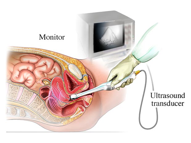
Transvaginal Ultrasound

Preparation for transvaginal ultrasound
Typically, no special preparation is required for this ultrasound, but some doctors may recommend emptying the bladder before the procedure. This helps improve image quality. Additionally, the patient should wear loose, comfortable clothing and may need to remove the lower-body garments.
Uses of transvaginal ultrasound
- Early pregnancy evaluation: To confirm pregnancy, assess fetal health, and diagnose ectopic or abnormal pregnancies.
- Assessment of pelvic pain: To identify the cause of sudden or chronic pelvic pain.
- Diagnosis of menstrual issues: Such as abnormal or irregular bleeding.
- Detection of ovarian cysts or masses: For diagnosing cysts, fibroids, or tumors in the ovaries and uterus.
- Post-surgical pelvic evaluation: To assess the success of pelvic surgeries or check for infections.
Procedure for transvaginal ultrasound
The doctor or technician gently inserts the ultrasound transducer, which is covered with a lubricating gel, into the patient's vagina. The transducer sends images to the ultrasound machine, which produces clear images of the internal pelvic organs. This process usually takes a few minutes and may cause some discomfort but is generally not painful.
Benefits of transvaginal ultrasound
High accuracy: This ultrasound allows for more precise evaluation by the specialist, as the vaginal probe enables detailed examination of internal organs, follicle count and size, endometrial thickness, causes of abnormal bleeding, and more.
Non-invasive method: This is a non-invasive procedure that does not require surgery or exposure to harmful radiation.
Short duration: The ultrasound usually takes a short amount of time, and the patient can quickly return to their daily activities afterward.
